Archives
Dolce Vita Hotel
E-Cat
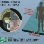
Elsevier’s Interactive Anatomy – Elbow Joint & Cubital Fossa
 |
|
| Category | Multimedia |
|---|---|
| Genre | Healthcare |
| Players | N/A |
| DVC | DVC required |
| Producer | Zoutewelle Multimedia |
| Publisher | Elsevier Science |
| Year | 1998 |
| Catalogue # | N/A |
| EAN | 0-444-82259-3 (Volume 2 Disk 3) |
| Discs | 1 |
| Videos | |
| Screenshots | |
| Covers |
|
| Controller | Remote |
| Description | Elbow Joint & Cubital Fossa is the third disc of the second volume of the most complete anatomical atlas available in electronic format – no printed medium can offer this amount of information. This volume covers the anatomy of the Upper Limb, which, next to the Elbow Joint & Cubital Fossa, contains the Shoulder Joint & Axilla and the Hand & Wrist. Elbow Joint & Cubital Fossa offers you more than 9,000 images of normal anatomy and 1,200 correlative images from CT, MRI and histology, and enables medical professionals – especially hand surgeons, plastic and reconstructive surgeons, traumatologists, general and orthopedic surgeons, radiologists, anatomists and neurosurgeons – to explore interactively the major structures of the humeroulnar, humeroradial and proximal radioulnar joints, the common flexor and extensor attachments, the detailed topography of deep and superficial radial, median and ulnar nerves, and the brachial artery and collateral vessels. You will be able to trace the major cubital nerves and blood vessels as they pass the elbow joint, and follow the path of ulnar and median nerves. It provides a unique view of structures, to be studied in the three cardinal planes. Elbow Joint & Cubital Fossa also includes Mallory-Cason stained histological images in the axial plane from the same elbow joint and cubital fossa region. All structures are displayed as continuous cross-sectional photographs in coronal, sagittal and axial directions, all within the same tissue block. Cross-sections can be magnified up to 4 times, and anatomy, CT, MRI and histology can be viewed in a “split-screen” mode. These sophisticated, high density images can be displayed as stills or video, while the structure names (20,000 labels in each viewing direction) can be displayed with a click, not only in the normal anatomy view, but also in the enlarged view. You can also travel step-wise through the images. Labels are available in English and according to the latest Nomina Anatomica. Many features included in the disc – such as “split screen” mode, “zoom” function, labelling in the “zoom” function, structure names in English – are the result of continuous market research and user feedback. |
| Remarks | This disc is a CD-i Bridge, it will play on CD-i players and Windows 95 & 98. |
| Regional | |
| CD-i Emulator | |
| MAME | |
| Reviews | |
| Interviews | |
| Downloads | |
| Credits | |
Elsevier’s lnteractive Anatomy – Paranasal Sinuses & Anterior Skull Base
 |
|
| Category | Professional |
|---|---|
| Genre | Healthcare |
| Players | N/A |
| DVC | DVC required |
| Producer | Zoutewell Multimedia |
| Publisher | Elsevier Science |
| Year | 1994 |
| Catalogue # | N/A |
| EAN | 0-444-89985-5 (Volume 1 Disk 1) |
| Discs | 1 |
| Videos | |
| Screenshots | |
| Covers | |
| Controller | Remote |
| Description | PARANASAL SINUSES & ANTERIOR SKULL BASE is the first disc of the most complete ENT atlas of normal anatomy available in electronic format – no printed medium can offer this amount of information.This first disc presents 11,000 anatomical cross-sections and 700 correlative images from CT, MRI and histology, and will enable medical professionals – specifically ENT surgeons, radiologists, anatomists, maxillofacial surgeons, ophthalmologists and plastic surgeons – to play an interactive part in exploring the major structures of the nose, paranasal sinuses, orbit, sella turcica, hypophysis, cavernous sinus, and sphenopalatine fossa. All structures are displayed as continuous cross-sectional photographs (taken from the same head of a healthy specimen) in coronal, saggital and axial directions. These sophisticated, high density images can be displayed as stills or video animation, and can be viewed with or without the names of the structures in nomina anatomica. Disc I also includes Mallory-stained histological images in coronal direction from the same head. |
| Remarks | This disc is a CD-i Bridge, it will play on CD-i players and Windows 95 & 98. |
| Regional | |
| CD-i Emulator | |
| MAME | |
| Reviews | |
| Interviews | |
| Downloads | |
| Credits | |
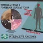
Elsevier’s lnteractive Anatomy – Temporal Bone & Posterior Cranial Fossa
 |
|
| Category | Professional |
|---|---|
| Genre | Healthcare |
| Players | N/A |
| DVC | DVC required |
| Producer | Zoutewelle Multimedia |
| Publisher | Elsevier Science |
| Year | 1994 |
| Catalogue # | N/A |
| EAN | 0-444-82016-7 (Volume 1 Disk 2) |
| Discs | 1 |
| Videos | |
| Screenshots | |
| Covers |
|
| Controller | Remote |
| Description | TEMPORAL BONE & POSTERIOR CRANIAL FOSSA is the second disc of the most complete ENT atlas available in electronic format – no printed medium can offer this amount of information. This second disc presents 9,000 images of normal anatomy and 1,200 correlative images from CT, MRI and histology, and enables medical professionals – specifically ENT surgeons, radiologists, anatomists, maxillofacial surgeons, and neurosurgeons – to explore interactively the major structures of the temporomandibular joint, tympanic cavity, labyrinth, facial canal, parapharyngeal space, and skull base (from sella turcica to foramen magnum). Disc II also includes Mallory-stained histological images in sagittal direction from the same head. All structures are displayed as continuous cross-sectional photographs in coronal, sagittal and axial directions. Cross-sections can be magnified up to 4 times, and anatomy, CT, MR and histology can be viewed in a “split screen” mode. These sophisticated, high density images can be displayed as stillsor video, and can be viewed either with or without the structure names (in English and/or nomina anatomica) – 25,000 name labels in total! New features included on this disc (“split screen” mode – “zoom” function – structurenames in English), are the result of extensive market research and user feedback. |
| Remarks | This disc is a CD-i Bridge, it will play on CD-i players and Windows 95 & 98. |
| Regional | |
| CD-i Emulator | |
| MAME | |
| Reviews | |
| Interviews | |
| Downloads | |
| Credits | |

